Visual Evoked Potentials (VEPs) Mechanism, Uses, and Benefits Behind this Vital Intraoperative Neuromonitoring Modality
DOI:
https://doi.org/10.5281/zenodo.10211280Keywords:
VEP, P100, ERG, visual evoked potentials, electroretinographyAbstract
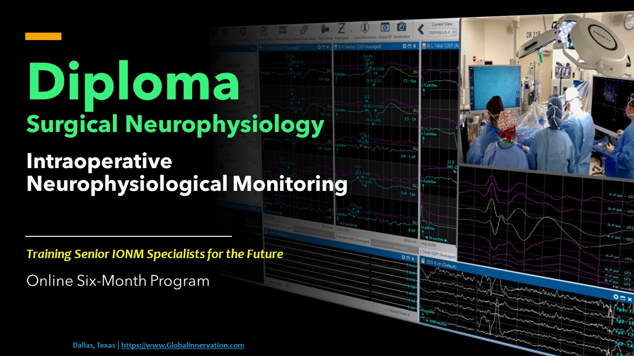
Visual evoked potential, or VEP is a neuromonitoring modality specifically used for evoking and recording neural signals in the visual/optic pathway – specifically, for detecting injuries surrounding the optic nerve and pre-chiasmatic areas. Visual Evoked Potentials (VEP), also known as Visual Evoked Response (VER), is a type of electrophysiological testing to measure the electrical potential resulting from a visual stimulus. VEP neuromonitoring is prescribed for individuals suffering from different visual pathologies such as multiple sclerosis, loss of vision, abnormalities in color vision, blurred or/and double vision, etc. There are two types of VEPs – pattern VEPs (PVEP) and flash VEPs (FVEP). Pattern VEPs is when the clinician asks the patient to watch a checkered pattern on a screen while recording optic neural signals from the brain. On the other hand, flash VEPs are almost the same type of test but with light-omitting goggles instead of presenting a visual pattern. Light-omitting goggles are placed on the eyes bilaterally, and responses are recorded from visual pathways, including the cornea, optic chiasm, and the occipital lobe (visual cortex). Both tests take no longer than an hour in duration in outpatient clinics. Some factors could influence the VEP responses, such as age, gender, or any medication taken before the test that could produce drowsiness in the patient; thus, it is important to follow instructions from the professional in charge of the neuromonitoring assessment.
References
Jahangiri FR, Ilyas S, Asdi R, Escobedo E, Seals M, “Protecting the Visual Pathways During Optic Nerve Surgery Using Intraoperative Visual Evoked Potentials (VEP).” Acta Scientific Clinical Case Reports 1.2 (2020): 01-06.
American Clinical Neurophysiology Society. “Guideline 9B: guidelines on visual evoked potentials”. American Journal of Electroneurodiagnostic Technology 46.3 (2006): 254-274. 2.
Jahangiri, FR. “Surgical Neurophysiology: An Introduction to Intraoperative Neurophysiological Monitoring (IONM)”. Second Edition. 193-95, April (2012).
Abulaban A and Jahangiri FR. “Establishment of Normal Visual Evoked Potentials (VEPs) Values in Adult Saudi Males”. Abstracts published in the proceeding of King Saud Bin Abdulaziz University for Health Sciences 15th Resident’s Day. Riyadh, Saudi Arabia: King Abdulaziz Medical City (2012): 36.
Thirumala PD, Habeych ME, Crammond DJ, Balzer JR. Neurophysiologic intraoperative monitoring of olfactory and optic nerves. J Clin Neurophysiol. 2011 Dec;28(6):538-42.
https://sydneynorthneurology.com.au/visual-evoked-potential-vep/
https://www.myvmc.com/investigations/visual-evoked-potential-vep/
Downloads
Published
How to Cite
Issue
Section
License
Copyright (c) 2023 J of Neurophysiological Monitoring

This work is licensed under a Creative Commons Attribution 4.0 International License.