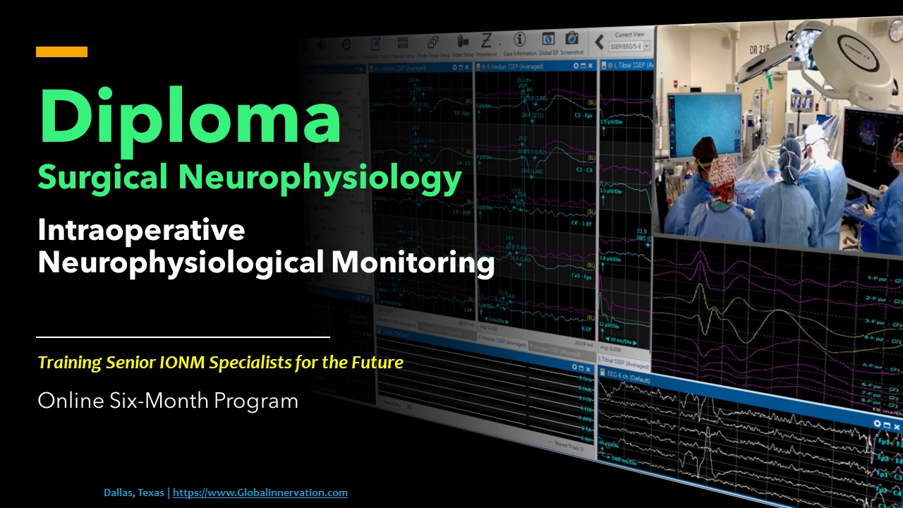
Spinal Cord Mapping with Somatosensory Evoked Potentials (SSEP) in Cervical and Thoracic Surgeries
DOI:
https://doi.org/10.5281/zenodo.10207942Keywords:
SSEP, MEP, EMG, mappin, cervical, thoracic, surgery, intramedullary tumor, phase reversalAbstract
Intraoperative neurophysiological monitoring (IONM) is commonly used in surgeries to reduce post-operative deficits. IONM uses multiple modalities, including SSEP, TCeMEP, and EMG. In this chapter, we will discuss the use of SSEP in mapping the spinal cord for cervical and thoracic surgeries that involve intramedullary tumor resections.
We use SSEP phase reversals and collision studies to find the midline raphe to make a safe incision. We can either directly stimulate the spinal cord and record from the brain to tell the difference between the right and left dorsal columns, or we can stimulate the peripheral nerve (median or tibial), cause a collision with an epidural stimulator, and look for decreased amplitude of signal in the cortex.
Mounting evidence suggests that mapping the spinal cord using SSEP is a safe and effective method that can be used to determine the location of the midline raphe separating the sensory tracts for the midline myelotomy and epidural stimulator placement. SSEP monitoring is also an effective way to prevent postoperative deficits by constantly monitoring SSEP throughout the surgery.
In conclusion, we can say that cortical spinal mapping helps locate the midline raphe, which can help surgeons during surgical procedures to help keep the dorsal columns unharmed.
References
Balzer, J. R., Tomycz, N. D., Crammond, D. J., Habeych, M., Thirumala, P. D., Urgo, L., & Moossy, J. J. (2011). Localization of cervical and cervicomedullary stimulation leads for pain treatment using median nerve somatosensory evoked potential collision testing. Journal of neurosurgery, 114(1), 200–205.
Biscevic M., Sehic A., Krupic F., (2020). Intraoperative neuromonitoring in spine deformity surgery: modalities, advantages, limitations, medicolegal issues – surgeons’ views. Spine, 5, 9-16.
Chen, Y., Wang, B. P., Yang, J., & Deng, Y. (2017). Neurophysiological monitoring of lumbar spinal nerve roots: A case report of postoperative deficit and literature review. International journal of surgery case reports, 30, 218–221.
Gertsch JH, Moreira JJ, Lee GR, Hastings JD, Ritzl E, Eccher MA, Cohen BA, Shils JL, McCaffrey MT, Balzer GK, Balzer JR, Boucharel W, Guo L, Hanson LL, Hemmer LB, Jahangiri FR, Mendez Vigil JA, Vogel RW, Wierzbowski LR, Wilent WB, Zuccaro JS, Yingling CD; membership of the ASNM. Practice guidelines for the supervising professional: intraoperative neurophysiological monitoring. J Clin Monit Comput. 2019 Apr; 33:175-183.
Mehta A.I., Mohrhaus C.A., Husain A.M., Karikari I.O., Hughes B., Hodges T., Gottfried O., Bagley C.A. (2012). Dorsal column mapping for intramedullary spinal cord tumor resection decreases dorsal column dysfunction. J Spinal Disord Tech. 25, 205-9.
Nair, D., Kumaraswamy, V. M., Braver, D., Kilbride, R. D., Borges, L. F., & Simon, M. V. (2014). Dorsal column mapping via phase reversal method: the refined technique and clinical applications. Neurosurgery, 74, 437–446.
Pajewski, T. N., Arlet, V., & Phillips, L. H. (2007). Current approach on spinal cord monitoring: the point of view of the neurologist, the anesthesiologist and the spine surgeon. European spine journal: official publication of the European Spine Society, the European Spinal Deformity Society, and the European Section of the Cervical Spine Research Society, 16 Suppl 2, S115–S129.
Quinones-Hinojosa, A., Gulati, M., Lyon, R., Gupta, N., & Yingling, C. (2002). Spinal Cord Mapping as an Adjunct for Resection of Intramedullary Tumors: Surgical Technique with Case Illustrations. Neurosurgery, 51, 1199-1207.
Simon M.V., Chiappa K.H., Borges L.F. (2012). Phase reversal of somatosensory evoked potentials triggered by gracilis tract stimulation: case report of a new technique for neurophysiologic dorsal column mapping. Neurosurgery. 70, E783-8.
Yanni D.S., Ulkatan S., Deletis V., Barrenechea I.J., Sen C., Perin N.I. (2010). Utility of neurophysiological monitoring using dorsal column mapping in intramedullary spinal cord surgery. Journal of neurosurgery 12, 623-8.
Downloads
Published
How to Cite
Issue
Section
License
Copyright (c) 2023 J of Neurophysiological Monitoring

This work is licensed under a Creative Commons Attribution 4.0 International License.


